How Our Technology is Revolutionising Traditional Dermatology
Why is Using Cutting-Edge Technology in Dermatology is So Important?
By using the latest technology in dermatology, we can sometimes make an immediate diagnosis, all whilst being pain-free and extremely reliable! This in turn allows our patients to receive the treatment they need faster.
We are leading consultants offering world-class dermatology that is painless, non-invasive, and extremely effective. Our cutting-edge technology combined with our internationally renowned team provides an exceptional standard of care to our patients. Our approach enables painless, non-invasive, and rapid results that save patients from the costs and scarring of unnecessary biopsies.
Other mole check services run by non-dermatologists may only provide a physical biopsy as a means of diagnosis. We find this approach to incur unnecessary scarring, costs, time, and anxiety for patients. What sets us apart from other skin clinics in the UK is that all skin examinations are carried out by one of our consultant dermatologists. Our mission is to revolutionise traditional dermatology by using the latest technology in dermatology, which ensures a better experience for our patients. However, if your dermatologist believes a procedure is appropriate, we do have a theatre on-site where your dermatologist will personally excise it and send it for histopathology.
The Latest Generation Dermatoscope for Painless & Effective Mole Checks
Digital dermoscopy is a hand-held instrument that our consultant dermatologists use to examine suspicious moles, lesions, as well as many other skin conditions. Dermoscopy is a noninvasive method that allows the in vivo evaluation of colors and microstructures of the epidermis, the dermoepidermal junction, and the papillary dermis not visible to the naked eye. These structures are specifically correlated to histologic features. In other words, the doctor can sometimes provide an immediate diagnosis without the need for a physical biopsy! We can therefore save our patients the time, scarring, costs, and anxiety associated with an unnecessary procedure.
Its incredible magnification power when placed on the skin gives the doctor a window into the skin. The images are digitally stored and compared with future consultations. The dermatoscope is safe for pregnant women, children, and even inflamed skin. By using the latest generation dermatoscope, your dermatologist will have an immediate idea as to the health of your skin. The dermatoscope produces results that are rapid, painless, and extremely reliable. This is how we ensure a better experience for our patients, as well as provide a definitive diagnosis for patients seeking a reliable second opinion.

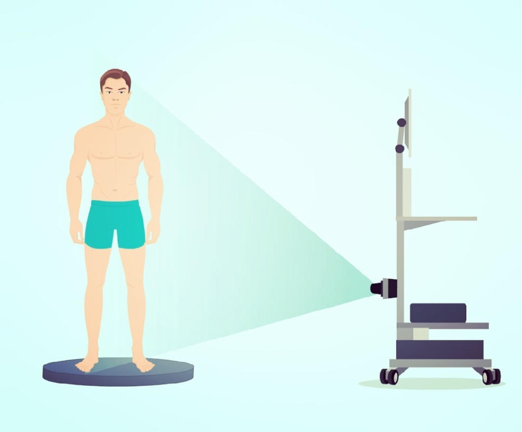
Total Body Mapping (TBM) to Track Your Moles over Time
You may also opt for total body mapping (TBM) at your mole check. 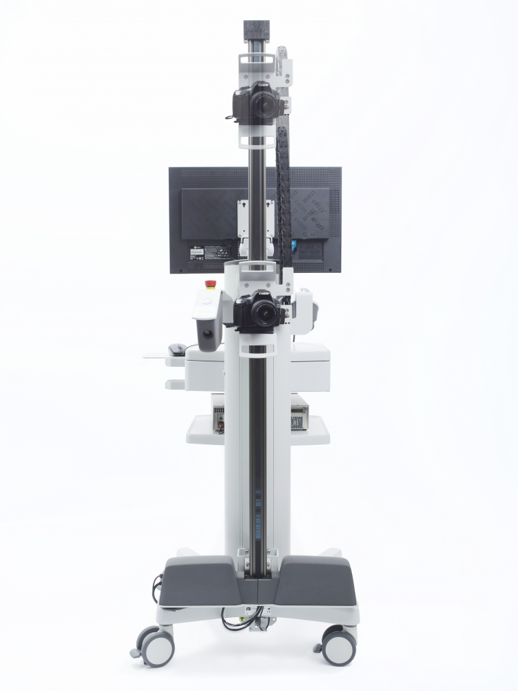 TBM is a remarkable machine that takes detailed images of the person’s entire body, thus creating an accurate record of their skin over time. The cutting-edge software in combination with our consultant dermatologists can detect any changes in moles at each annual mole check. This technology is ideal for people with a high number of freckles or moles, or who have had a previous skin cancer diagnosis. Annual TBM is the best way to keep your skin monitored for skin cancer. Melanoma and other skin cancers respond very well to treatment if detected at the early stages. Therefore, TBM can help catch changes as soon as possible, thus preventing mortality.

Benefit from our technology’s remarkable potential to take your time to know from weeks to minutes.
We give patients peace of mind knowing that their skin is being continuously monitored by leading dermatologists and the most advanced technology for dermatology in the UK. Book your highly specialised mole check today!
If you do have any skin worries, or notice any irregularities or new moles on your skin, it’s important to get them checked out promptly:
call us on +44 (0)20 3575 1474 or visit our contact page to get in touch as soon as possible.
References
- Salerni G, Terán T, Puig S, et al. Meta-analysis of digital dermoscopy follow-up of melanocytic skin lesions: a study on behalf of the International Dermoscopy Society. J Eur Acad Dermatol Venereol. 2013;27(7):805-814. doi:10.1111/jdv.12032
- Tschandl P. Sequential digital dermatoscopic imaging of patients with multiple atypical nevi. Dermatol Pract Concept. 2018;8(3):231-237. Published 2018 Jul 31. doi:10.5826/dpc.0803a16
- Salerni G, Carrera C, Lovatto L, et al. Benefits of total body photography and digital dermatoscopy (“two-step method of digital follow-up”) in the early diagnosis of melanoma in patients at high risk for melanoma. J Am Acad Dermatol. 2012;67(1):e17-e27. doi:10.1016/j.jaad.2011.04.008
- Malvehy J, Puig S. Follow-up of melanocytic skin lesions with digital total-body photography and digital dermoscopy: a two-step method. Clin Dermatol. 2002;20(3):297-304. doi:10.1016/s0738-081x(02)00220-1

Leave a Reply
Want to join the discussion?Feel free to contribute!